Leeds shoots for 100% digitisation of histopathology slides
- 16 October 2018
Leeds Teaching Hospitals NHS Trust and the University of Leeds have started digitally scanning all their pathology slides in a move to enable AI-supported diagnoses.
Some 1,000 slides are being scanned by Leeds Teaching Hospital every day using technology from medical device firm, Leica Biosystems.
The aim is to digitise 100% of Leeds Teaching Hospital’s histopathology slides so that they can be easily shared across multi-disciplinary teams, allowing for greater collaboration and better decision-making about patient treatment.
The programme also looks at the possibility of harnessing AI (artificial intelligence) to improve the speed and accuracy of diagnoses: by feeding the images into a machine learning algorithm, a computer could be trained to spot patterns that may be missed by traditional methods.
For example, computers could help to detect small areas of cancer that have spread to lymph nodes, better calculate and measure tumours, and quickly flag abnormalities tissue samples to pathologists.
Dr Darren Treanor, a consultant pathologist at Leeds Teaching Hospitals NHS Trust and University of Leeds, said: “The way we are digitising pathology services in Leeds enables us to produce the quality images pathologists need to diagnose patients quickly and safely.
“Going digital not only allows the pathologists to do their work on a computer, but it also unlocks the possibility of using the computer to help make the diagnosis, offering huge potential for the future.”
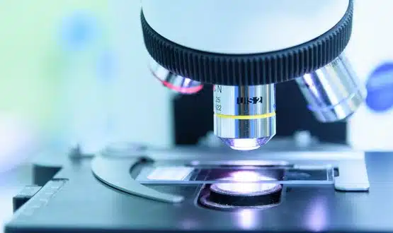
The traditional approach used by pathologists to diagnose cancer is to examine glass slides under a microscope. This throws up a multitude of challenges around the way slides are stored, shared and transported, resulting in costly solutions.
‘Digital pathology’ overcomes these challenges by making processes electronic, allowing pathologists to more easily work off computers.
At Leeds Teaching Hospitals, digital slides are created by feeding glass slides into a Leica Biosystems scanner, which produces an image of the sample at a resolution of 200,000 dots per inch.
Over a thousand slides are prepared and scanned by the trust daily, creating one terabyte of new data every day. It claimed that, if all the glass slides produced in the histopathology department in one month were laid out end to end, it would stretch for a mile.
Leeds Teaching Hospitals has so far scanned 200,000 slides containing patient tissue samples. Meanwhile, the University of Leeds houses an additional 386,000 anonymised digital slides for research and teaching purposes.
By March 2019, it is expected that all pathologists at Leeds Teaching Hospitals will have been validated to diagnose from digital images.
Trust programme manager, Basharat Hussain, said demand for pathology services would continue to increase and that digital pathology had the potential to allowed resources to be pooled across hospitals.
Melissa Aquino, president of Leica Biosystems, added: “We invest in innovations which enable clinicians to rapidly and efficiently provide a highly confident diagnosis. The mainstream adoption of digital pathology fits squarely within that framework. We are proud to have worked with the Leeds team and enabled 100% digitisation of their histopathology slides.”
The digital pathology programme is backed by a £1m grant from local charity, Leeds Cares.
Leeds Teaching Hospitals suffered a major IT blowout in its pathology department in September 2016, which a subsequent review deemed to be the result of human error.
A spokesperson said the digital pathology programme was not linked to the incident.